Periodontal EDS
Last reviewed: 2026-06-09
Periodontal Ehlers-Danlos Syndrome (pEDS) is a rare connective tissue disorder caused by changes in the C1R or C1S gene. Although these genes are part of the complement system, in periodontal EDS the changes affect how connective tissue is organized, particularly in the gums and skin. Its defining feature is severe, early-onset periodontitis that causes the gums to recede and can lead to early loss of teeth. People with pEDS also frequently have pretibial plaques (firm patches on the shins), joint hypermobility, and stretchy, fragile skin that scars abnormally. pEDS is inherited in an autosomal dominant pattern, meaning a change in one copy of the gene is enough to cause the condition.
Genetics & Inheritance
C1R or C1S
Caused by mutations at specific locations in ONE copy of C1R or C1S
Signs & Features of pEDS
Severe, early-onset periodontitis
Periodontitis is inflammation and breakdown of the tissues that hold the teeth in place, including the gums and the bone beneath. In periodontal EDS it is severe and begins unusually early in life, reflecting the weakness of these supporting connective tissues. It is the defining feature of this type.

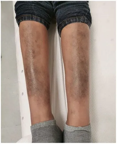
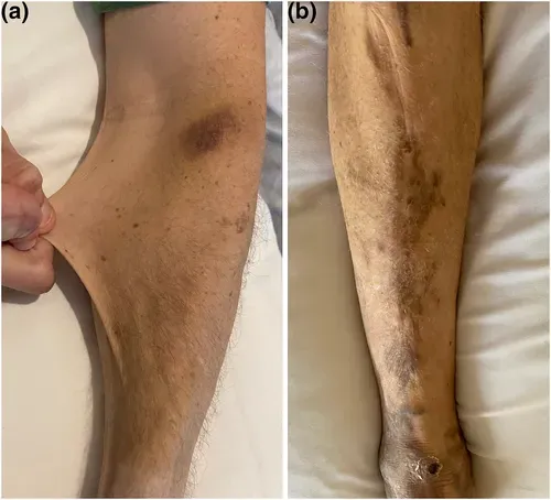
Pretibial plaques
Pretibial plaques are firm, discolored, slightly raised patches of skin over the front of the shins. They are a recognized feature of periodontal EDS and tend to form over areas prone to knocks and pressure. They reflect changes in the skin and its underlying tissue in this type.
Joint hypermobility
Joints that move well beyond the normal range, affecting many joints throughout the body rather than just one or two. In the rare EDS types this is present from birth and is often severe, contributing to joint instability, pain, and frequent sprains.
Bone marrow abnormalities
Bone marrow is the tissue inside bones where blood cells are made. Changes affecting the bone marrow have been reported in some people with periodontal EDS. How common or how significant these changes are is still being worked out by researchers.
Stretchy, fragile skin
Hyperextensible skin stretches further than usual when pulled and then springs back into place, rather than staying loose. It reflects changes in the collagen that gives skin its strength and elasticity, and is a recognized feature across many of the EDS types. The degree of stretch varies from mild to pronounced.
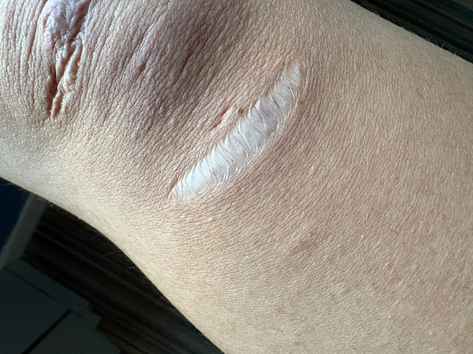
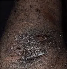
Atrophic scars
Atrophic scars are wide, thin, sunken scars that form when fragile skin heals poorly after injury. In several rare EDS types, even minor wounds can leave papery, wrinkled scars — often compared to cigarette paper — most visibly over the forehead, knees, elbows, and shins.
Recessing gums
Gingival recession is when the gums pull back from the teeth, exposing more of the tooth and its root. In periodontal EDS it occurs alongside the breakdown of the tissues that support the teeth. It reflects the fragility of these gum tissues in this type.
Photos are illustrative examples of individual findings; appearance varies widely from person to person. This page is educational and is not a diagnostic tool — see our disclaimer.
Sources
Brady AF, Demirdas S, Fournel-Gigleux S, Ghali N, Giunta C, Kapferer-Seebacher I, et al. The Ehlers-Danlos syndromes, rare types. Am J Med Genet C Semin Med Genet. 2017;175(1):70-115.
Malfait F, Francomano C, Byers P, Belmont J, Berglund B, Black J, et al. The 2017 international classification of the Ehlers-Danlos syndromes. Am J Med Genet C Semin Med Genet. 2017;175(1):8-26.
Angwin C, Ghali N, et al. Ehlers-Danlos syndromes, rare types: Clinical and molecular review. Eur J Hum Genet. 2023;31(2):131-142.
Living with Periodontal EDS? You're not alone.
Join Our Community