Classical EDS
Last reviewed: 2026-06-09
Classical Ehlers-Danlos Syndrome (cEDS) is a connective tissue disorder caused by changes in the COL5A1, COL5A2, or COL1A1 gene, which affect type V (or type I) collagen. Its hallmark features are highly stretchy skin, severe generalized joint hypermobility, and significant wound healing complications, including widened, atrophic scarring and delayed healing. People with cEDS also commonly have easy bruising with hemosiderin staining, skin fragility, recurrent subluxations and dislocations, hernias, and piezogenic papules. cEDS is inherited in an autosomal dominant pattern, meaning a change in one copy of the gene is enough to cause the condition.
Genetics & Inheritance
COL5A1, COL5A2, or COL1A1
Caused by mutations at specific locations in ONE copy of COL5A1, COL5A2, or COL1A1
Signs & Features of cEDS
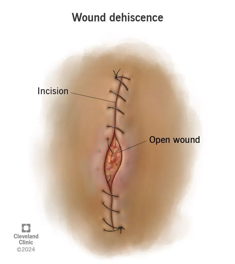
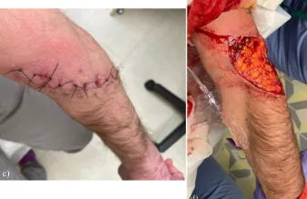
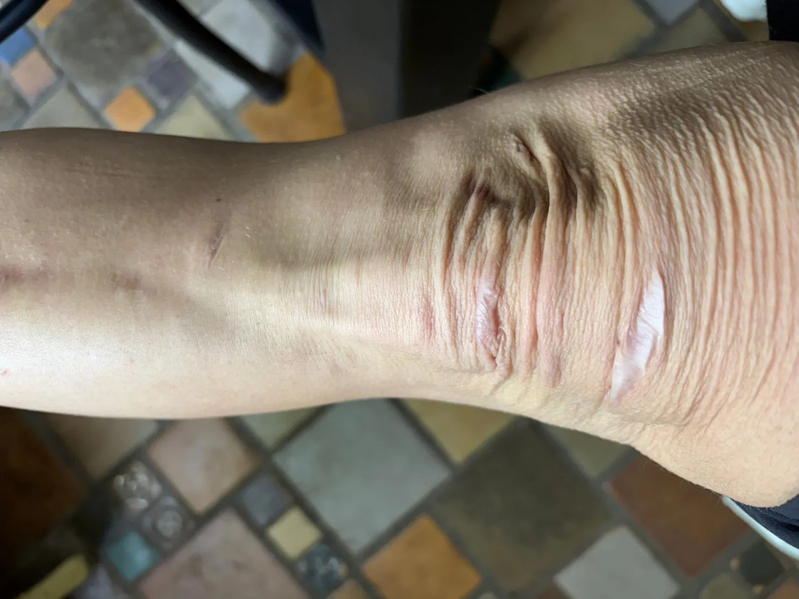
Significant wound healing complications like dehiscence, severe atrophic scarring, and failure to progress through stages of wound healing
In classical EDS, wounds often heal slowly and poorly. Healing edges may pull apart again (dehiscence), and the resulting scars are frequently wide, thin, and sunken (atrophic). These difficulties stem from the fragile skin and altered collagen that make it hard for a wound to knit together strongly.
Severe generalized joint hypermobility
Joints that move well beyond the normal range, affecting many joints throughout the body rather than just one or two. In the rare EDS types this is present from birth and is often severe, contributing to joint instability, pain, and frequent sprains.
Subluxations/dislocations
A subluxation is a partial slip of a joint out of position, while a dislocation is a complete one. Because the connective tissue that normally holds joints in place is weakened in several rare EDS types, these slips can happen easily and repeatedly, sometimes with little or no force. Over time they contribute to joint pain and instability.
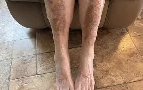
Easy bruising with hemosiderin staining
When bruises occur repeatedly in the same area, iron released from the blood (hemosiderin) can build up and leave a lasting brown or rust-colored discoloration of the skin. This is often seen over the shins and other areas prone to knocks in classical EDS. It reflects both the easy bruising and the slow clearance of repeated bleeding under fragile skin.
Bilateral piezogenic papules
Piezogenic papules are small soft bumps that appear on the heels or wrists when weight or pressure is put on them, caused by fat pushing through gaps in the surrounding connective tissue. They often disappear when the pressure is released. They are common in classical EDS and reflect the laxity of the tissue that normally keeps the fat in place.
Skin fragility
Fragile skin tears, splits, or breaks more easily than usual, sometimes from minor knocks or friction. In several rare EDS types the skin is mechanically weak because its supporting collagen is altered, so wounds can be larger or deeper than the injury would suggest. This fragility also makes wounds harder to close and slower to heal.
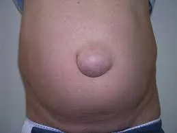
Hernia(s)
A hernia is a bulge that forms when an internal organ or tissue pushes through a weak spot in the muscle or connective tissue wall meant to contain it. Common sites include the navel (umbilical) and groin. In rare EDS types the supporting tissue is weaker than usual, so hernias can form more readily and sometimes recur.
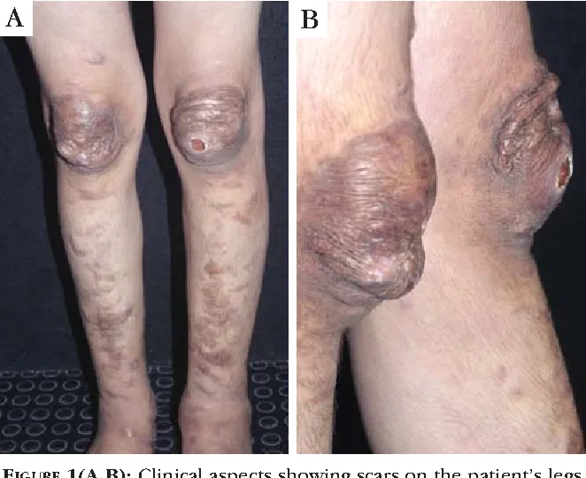
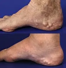
Subcutaneous spheroids
Subcutaneous spheroids are small, firm, pea-like lumps that can be felt under the skin, often over the forearms and shins. They are thought to be small areas of fat that have lost their blood supply and become firm or calcified. They are a recognized feature of classical EDS and are usually painless.
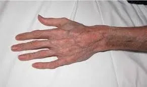
Acrogeria
Acrogeria refers to an aged appearance of the hands and feet, where the skin looks thin, and the underlying veins and tendons are unusually visible. It results from reduced fatty padding and thin skin over these areas. It is most characteristic of vascular EDS, though it is described in other rare types as well.
Redundant skin on the joints
Redundant skin is extra, loose skin that gathers in folds, often most noticeable over joints such as the knuckles, elbows, and knees. It reflects the increased laxity and stretchiness of the skin in several rare EDS types. The folds may be more apparent when the joint is relaxed.
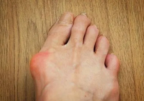
Hallux Valgus
Hallux valgus, commonly called a bunion, is when the big toe angles inward toward the other toes and a bony bump forms at its base. In rare EDS types the lax ligaments of the foot allow the joint to drift out of alignment more easily. It can affect the shape and comfort of the foot.
Scoliosis
These terms describe curves of the spine: scoliosis is a sideways curve, kyphosis an outward (rounding) curve, and lordosis an inward (sway) curve. In the rare EDS types the ligaments and muscles supporting the spine are lax, so the spine is less well held in alignment and curves can develop, sometimes in early life. The degree of curvature ranges widely between people.
Organ prolapse
Prolapse is when an internal organ slips downward from its usual position because the connective tissue and muscle supporting it are weakened. It can affect organs such as the bowel, bladder, uterus, or rectum. In rare EDS types the supporting tissue is more lax, which can make prolapse more likely.
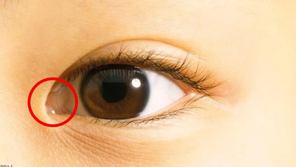
Epicanthal folds
Epicanthal folds are small folds of skin that run from the upper eyelid toward the inner corner of the eye, partly covering it. They are a normal variation in many people and are also noted as a facial feature in some rare EDS types. They are simply a characteristic of how the eyelid skin is shaped.
Photos are illustrative examples of individual findings; appearance varies widely from person to person. This page is educational and is not a diagnostic tool — see our disclaimer.
Sources
Brady AF, Demirdas S, Fournel-Gigleux S, Ghali N, Giunta C, Kapferer-Seebacher I, et al. The Ehlers-Danlos syndromes, rare types. Am J Med Genet C Semin Med Genet. 2017;175(1):70-115.
Malfait F, Francomano C, Byers P, Belmont J, Berglund B, Black J, et al. The 2017 international classification of the Ehlers-Danlos syndromes. Am J Med Genet C Semin Med Genet. 2017;175(1):8-26.
Angwin C, Ghali N, et al. Ehlers-Danlos syndromes, rare types: Clinical and molecular review. Eur J Hum Genet. 2023;31(2):131-142.
Living with Classical EDS? You're not alone.
Join Our Community