Arthrochalasia EDS
Last reviewed: 2026-06-09
Arthrochalasia Ehlers-Danlos Syndrome (aEDS) is a very rare connective tissue disorder caused by changes in the COL1A1 or COL1A2 gene, which affect how type I collagen is processed in the body. Its most recognizable feature is congenital bilateral hip dislocation, present from birth, along with severe generalized joint hypermobility and recurrent subluxations and dislocations. People with aEDS often have loose, stretchy, thin-feeling skin, skin fragility, curvatures of the spine, and low muscle tone (hypotonia). aEDS is inherited in an autosomal dominant pattern, meaning a change in one copy of the gene is enough to cause the condition.
Genetics & Inheritance
COL1A1 or COL1A2
Caused by mutations at specific locations in ONE copy of COL1A1 or COL1A2
Signs & Features of aEDS
Congenital bilateral hip dislocation
A dislocated hip is one where the ball of the thigh bone sits outside its socket. In arthrochalasia EDS this is typically present in both hips from birth, reflecting the very loose joints and connective tissue that are a hallmark of the type. It is one of the features that often draws attention to the condition in the newborn period.
Severe generalized joint hypermobility
Joints that move well beyond the normal range, affecting many joints throughout the body rather than just one or two. In the rare EDS types this is present from birth and is often severe, contributing to joint instability, pain, and frequent sprains.
Subluxations and dislocations
A subluxation is a partial slip of a joint out of position, while a dislocation is a complete one. Because the connective tissue that normally holds joints in place is weakened in several rare EDS types, these slips can happen easily and repeatedly, sometimes with little or no force. Over time they contribute to joint pain and instability.
Loose, stretchy, thin feeling skin
Hyperextensible skin stretches further than usual when pulled and then springs back into place, rather than staying loose. It reflects changes in the collagen that gives skin its strength and elasticity, and is a recognized feature across many of the EDS types. The degree of stretch varies from mild to pronounced.
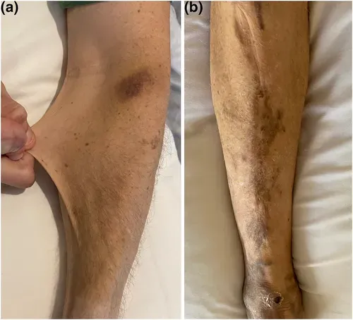
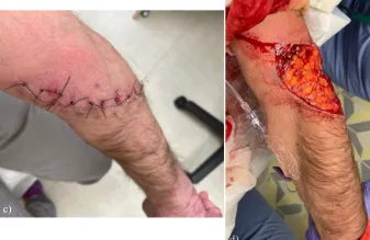
Skin fragility
Fragile skin tears, splits, or breaks more easily than usual, sometimes from minor knocks or friction. In several rare EDS types the skin is mechanically weak because its supporting collagen is altered, so wounds can be larger or deeper than the injury would suggest. This fragility also makes wounds harder to close and slower to heal.
Curvatures of the spine
These terms describe curves of the spine: scoliosis is a sideways curve, kyphosis an outward (rounding) curve, and lordosis an inward (sway) curve. In the rare EDS types the ligaments and muscles supporting the spine are lax, so the spine is less well held in alignment and curves can develop, sometimes in early life. The degree of curvature ranges widely between people.
Bone fragility
Fragile bones break more easily than expected, sometimes after minor falls or knocks. In types such as arthrochalasia EDS the collagen framework that gives bone part of its strength is affected, lowering bone resilience. This can show up as fractures earlier in life than usual.
Hypotonia
Hypotonia means reduced resting tension in the muscles, so the body can feel floppy and joints offer less natural resistance to movement. In infants it may be noticed as difficulty with head control or a loose, relaxed posture. It is a common early feature of several rare EDS types and can contribute to delays in reaching motor milestones.
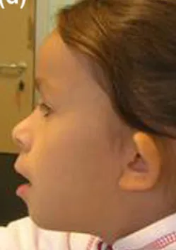
Craniofacial features
Facial differences are sometimes reported in arthrochalasia EDS, but they vary considerably from person to person and do not form a single, sharply defined pattern. When present, they may include subtle differences in the shape of the skull and the proportions of the face, which can be more noticeable in infancy. They are not in themselves a defining feature of the type.
Motor delay
Motor delay means reaching physical milestones such as sitting, crawling, or walking later than usual. In the rare EDS types it often reflects a combination of low muscle tone and very loose joints, which make stable, coordinated movement harder to achieve. The pattern and degree of delay differ from child to child.
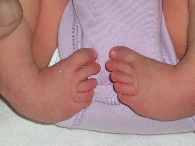
Foot deformities
Several rare EDS types are associated with differences in the shape or position of the feet, present from birth or developing over time. One example is club foot (talipes equinovarus), in which the foot is turned inward and downward. These differences arise from the lax connective tissue and altered muscle balance around the developing foot.
Photos are illustrative examples of individual findings; appearance varies widely from person to person. This page is educational and is not a diagnostic tool — see our disclaimer.
Sources
Brady AF, Demirdas S, Fournel-Gigleux S, Ghali N, Giunta C, Kapferer-Seebacher I, et al. The Ehlers-Danlos syndromes, rare types. Am J Med Genet C Semin Med Genet. 2017;175(1):70-115.
Malfait F, Francomano C, Byers P, Belmont J, Berglund B, Black J, et al. The 2017 international classification of the Ehlers-Danlos syndromes. Am J Med Genet C Semin Med Genet. 2017;175(1):8-26.
Angwin C, Ghali N, et al. Ehlers-Danlos syndromes, rare types: Clinical and molecular review. Eur J Hum Genet. 2023;31(2):131-142.
Living with Arthrochalasia EDS? You're not alone.
Join Our Community