Kyphoscoliotic EDS
Last reviewed: 2026-06-09
Kyphoscoliotic Ehlers-Danlos Syndrome (kEDS) is a rare connective tissue disorder caused by changes in the PLOD1 or FKBP14 gene, which affect how collagen is cross-linked and folded. Its defining feature is severe, congenital kyphoscoliosis (curvature of the spine) that worsens over time, along with marked muscle hypotonia at birth and generalized joint hypermobility. People with kEDS may also have scleral and ocular fragility, soft skin with widened scarring, arterial aneurysm, and a marfanoid build. kEDS is inherited in an autosomal recessive pattern, so a person must inherit a change in both copies of the gene to be affected.
Genetics & Inheritance
PLOD1 or FKBP14
Caused by mutations at specific locations in BOTH copies of PLOD1 or FKBP14
Signs & Features of kEDS
Severe kyphoscoliosis
Kyphoscoliosis is a combined curving of the spine both sideways (scoliosis) and forward (kyphosis). In kyphoscoliotic EDS it is typically present from birth or early infancy and tends to progress, because the muscles and ligaments supporting the spine are very lax. It is the defining feature of this type.

Muscle hypotonia
Hypotonia means reduced resting tension in the muscles, so the body can feel floppy and joints offer less natural resistance to movement. In infants it may be noticed as difficulty with head control or a loose, relaxed posture. It is a common early feature of several rare EDS types and can contribute to delays in reaching motor milestones.
Joint hypermobility
Joints that move well beyond the normal range, affecting many joints throughout the body rather than just one or two. In the rare EDS types this is present from birth and is often severe, contributing to joint instability, pain, and frequent sprains.
Eye involvement/scleral and ocular fragility
Ocular fragility means the outer tissues of the eye, including the white sclera, are weaker and more easily injured than usual. This reflects the altered connective tissue that gives the eye its structure in certain rare EDS types. It is part of the broader tissue fragility seen in these conditions.
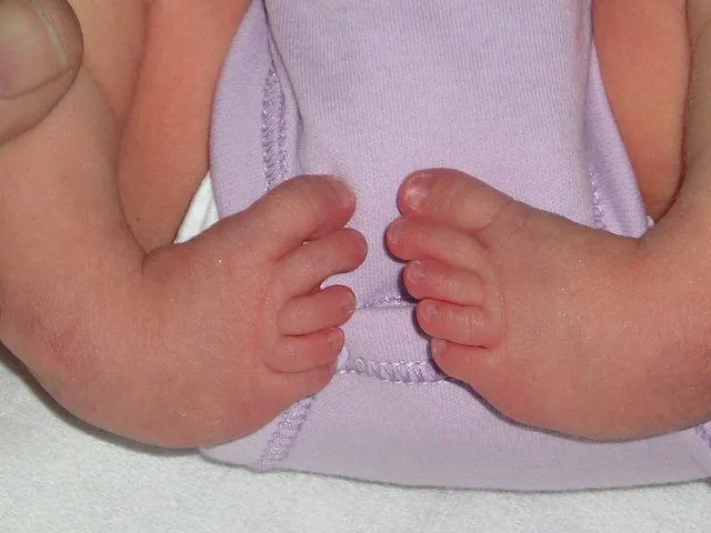
Head and foot deformities
Several rare EDS types are associated with differences in the shape or position of the feet, present from birth or developing over time. One example is club foot (talipes equinovarus), in which the foot is turned inward and downward. These differences arise from the lax connective tissue and altered muscle balance around the developing foot.
Severity, soft, doughy skin with widened scarring
Soft, doughy skin feels unusually soft and gives way easily to the touch, a little like dough. It is described in several rare EDS types and reflects changes in the texture and support of the skin. The skin may also be stretchy or fragile alongside this softness.
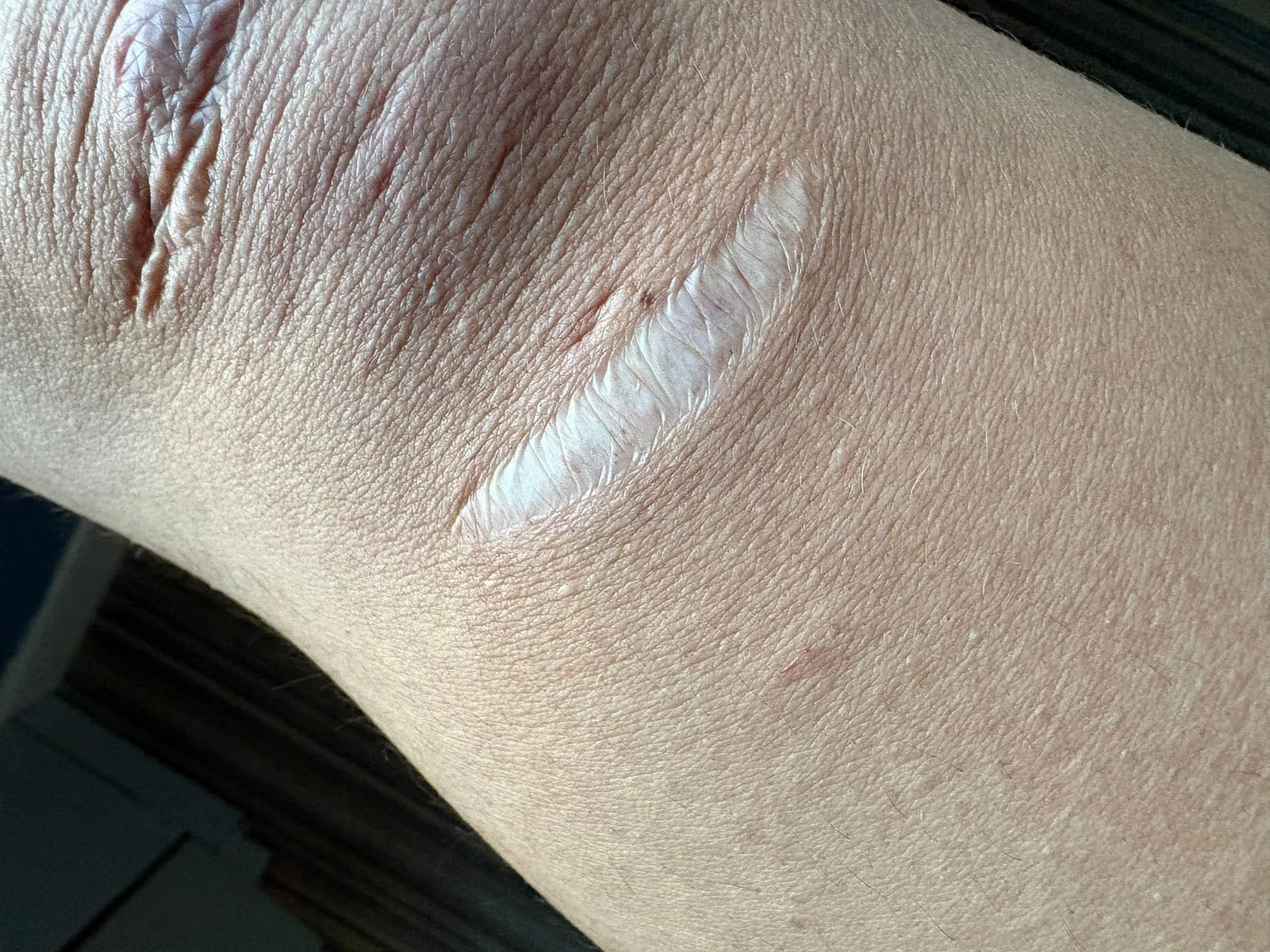
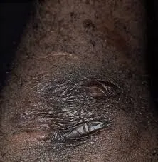
Atrophic scarring
Atrophic scars are wide, thin, sunken scars that form when fragile skin heals poorly after injury. In several rare EDS types, even minor wounds can leave papery, wrinkled scars — often compared to cigarette paper — most visibly over the forehead, knees, elbows, and shins.
Arterial aneurysm
An aneurysm is a bulge that forms in the wall of an artery where it has become weakened. In some rare EDS types the connective tissue in artery walls is altered, which can make these bulges more likely to form. It is a recognized feature that reflects the involvement of blood vessels in these conditions.
Craniofacial features
Kyphoscoliotic EDS can be associated with subtle facial characteristics. Reported features include a blue tint to the whites of the eyes and a degree of facial asymmetry that can develop alongside the spinal curvature. These features are mild and vary between individuals.
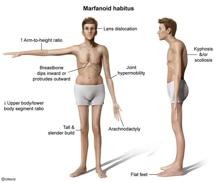
Marfanoid habitus
Marfanoid habitus describes a tall, slender build with long limbs, fingers, and toes relative to the body. It is seen in several connective tissue conditions, including some rare EDS types, and reflects the way altered connective tissue affects growth and proportion. The term describes a body shape rather than a single measurement.
Osteopenia/osteoporosis
Osteopenia and osteoporosis describe bone that is less dense than usual, with osteoporosis being the more marked. Lower bone density makes bones more prone to fracture. These features are reported in several rare EDS types, reflecting the role connective tissue plays in maintaining bone strength.
Photos are illustrative examples of individual findings; appearance varies widely from person to person. This page is educational and is not a diagnostic tool — see our disclaimer.
Sources
Brady AF, Demirdas S, Fournel-Gigleux S, Ghali N, Giunta C, Kapferer-Seebacher I, et al. The Ehlers-Danlos syndromes, rare types. Am J Med Genet C Semin Med Genet. 2017;175(1):70-115.
Malfait F, Francomano C, Byers P, Belmont J, Berglund B, Black J, et al. The 2017 international classification of the Ehlers-Danlos syndromes. Am J Med Genet C Semin Med Genet. 2017;175(1):8-26.
Angwin C, Ghali N, et al. Ehlers-Danlos syndromes, rare types: Clinical and molecular review. Eur J Hum Genet. 2023;31(2):131-142.
Living with Kyphoscoliotic EDS? You're not alone.
Join Our Community