Brittle Cornea Syndrome
Last reviewed: 2026-06-09
Brittle Cornea Syndrome (BCS) is a rare form of Ehlers-Danlos Syndrome caused by changes in the ZNF469 or PRDM5 gene, which disrupt the connective tissue that supports the eye. Its hallmark feature is a thin, fragile cornea that can tear or rupture after minor trauma, along with keratoconus or keratoglobus, blue sclera, and high myopia. People with BCS may also have hearing loss, small joint hypermobility, hip dysplasia, and a marfanoid build. BCS is inherited in an autosomal recessive pattern: a person must inherit a change in both copies of the gene to be affected.
Genetics & Inheritance
ZNF469 or PRDM5
Caused by mutations at specific locations in BOTH copies of ZNF469 or PRDM5
Signs & Features of BCS
Thin, fragile cornea
The cornea is the clear front window of the eye. In Brittle Cornea Syndrome it is thinner than usual, which makes it mechanically weaker and more easily injured. This thinning is a defining feature of the condition and underlies several of its other eye findings.
Ocular rupture
Ocular rupture is a tear of the outer wall of the eye, which can happen after a relatively minor injury when that wall is unusually thin and fragile, as in Brittle Cornea Syndrome. It is one of the more serious consequences of the weakened cornea and sclera seen in this condition. It reflects how the eye’s structural strength is reduced in this type.
Keratoconus/keratoglobus
Keratoconus and keratoglobus are changes in the shape of the cornea: in keratoconus it bulges into a cone, and in keratoglobus it becomes globe-shaped and thinned across its whole surface. Both arise when the cornea is structurally weak, as it is in Brittle Cornea Syndrome, and both can blur or distort vision. They reflect the same underlying corneal fragility seen in this type.
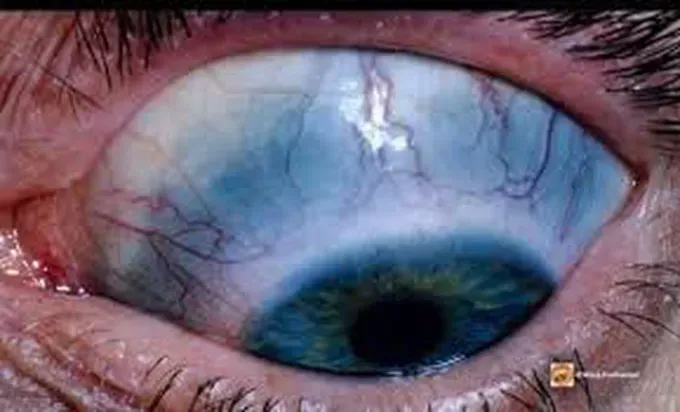
Blue sclera
The sclera is the white outer layer of the eye. When it is thinner than usual — as in Brittle Cornea Syndrome — the darker tissue underneath shows through, giving the whites of the eyes a blue or blue-grey tint.
High myopia
Myopia, or short-sightedness, means distant objects appear blurred. High myopia is a marked degree of this and is a recognized feature of Brittle Cornea Syndrome, where the shape and structure of the eye are altered. It reflects the same connective tissue changes that affect the cornea and sclera.
Hearing loss
Some rare EDS types, including Brittle Cornea Syndrome, are associated with reduced hearing. This can arise because connective tissue contributes to the structures of the ear that carry and process sound. The degree and type of hearing change vary between people.
Craniofacial features
Brittle Cornea Syndrome can be associated with subtle differences in the shape of the face and head. Reported features include prominent or deep-set eyes and a long, narrow facial appearance. These characteristics are mild and not present in everyone with the condition.
Small joint hypermobility
In some rare EDS types the extra range of movement is most noticeable in the small joints, such as those of the fingers and toes, rather than the large joints. This reflects the laxity of the ligaments that normally limit how far these joints bend. It can make the small joints prone to slipping or overextending.
Hip dysplasia
Hip dysplasia describes a hip socket that is shallow or poorly shaped, so the joint fits together less securely. In rare EDS types the lax connective tissue around the developing hip can contribute to this looser fit. It can make the hip more prone to instability.
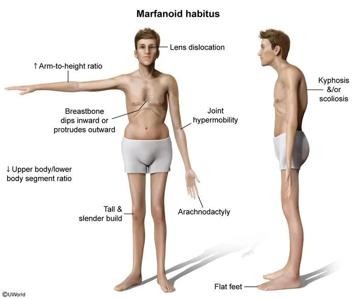
Marfanoid habitus
Marfanoid habitus describes a tall, slender build with long limbs, fingers, and toes relative to the body. It is seen in several connective tissue conditions, including some rare EDS types, and reflects the way altered connective tissue affects growth and proportion. The term describes a body shape rather than a single measurement.
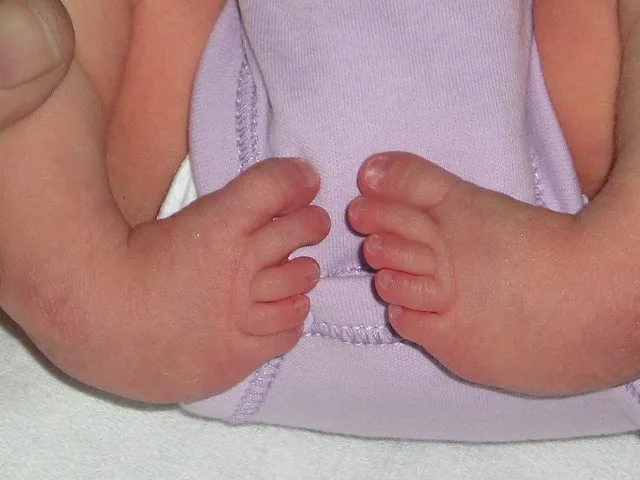
Foot deformities
Several rare EDS types are associated with differences in the shape or position of the feet, present from birth or developing over time. One example is club foot (talipes equinovarus), in which the foot is turned inward and downward. These differences arise from the lax connective tissue and altered muscle balance around the developing foot.
Scoliosis, kyphosis, and/or lordosis
These terms describe curves of the spine: scoliosis is a sideways curve, kyphosis an outward (rounding) curve, and lordosis an inward (sway) curve. In the rare EDS types the ligaments and muscles supporting the spine are lax, so the spine is less well held in alignment and curves can develop, sometimes in early life. The degree of curvature ranges widely between people.
Photos are illustrative examples of individual findings; appearance varies widely from person to person. This page is educational and is not a diagnostic tool — see our disclaimer.
Sources
Brady AF, Demirdas S, Fournel-Gigleux S, Ghali N, Giunta C, Kapferer-Seebacher I, et al. The Ehlers-Danlos syndromes, rare types. Am J Med Genet C Semin Med Genet. 2017;175(1):70-115.
Malfait F, Francomano C, Byers P, Belmont J, Berglund B, Black J, et al. The 2017 international classification of the Ehlers-Danlos syndromes. Am J Med Genet C Semin Med Genet. 2017;175(1):8-26.
Angwin C, Ghali N, et al. Ehlers-Danlos syndromes, rare types: Clinical and molecular review. Eur J Hum Genet. 2023;31(2):131-142.
Living with Brittle Cornea Syndrome? You're not alone.
Join Our Community